Experimental Evidence of Functional Muscle Modulation

Endopeel Physiology

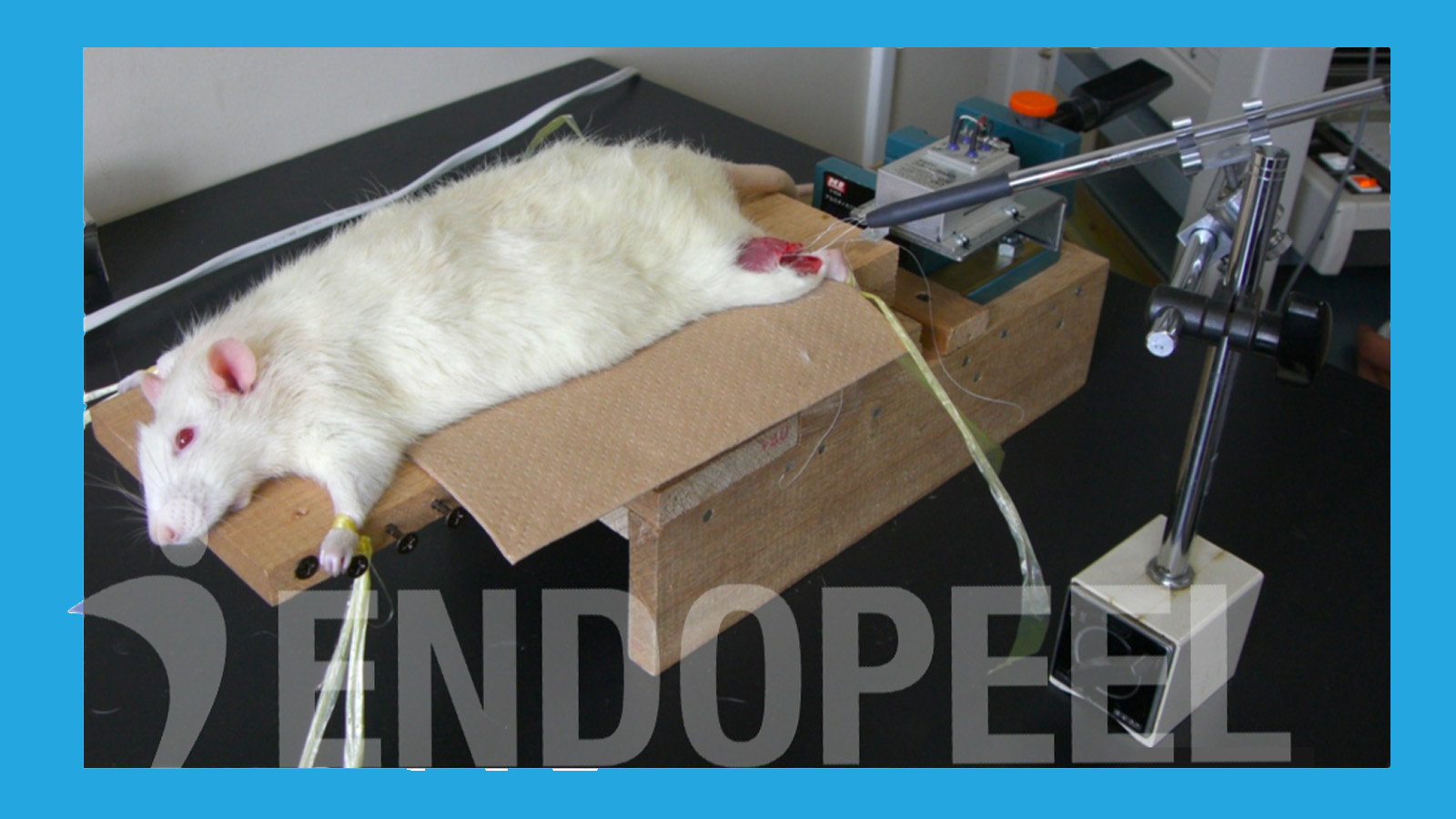
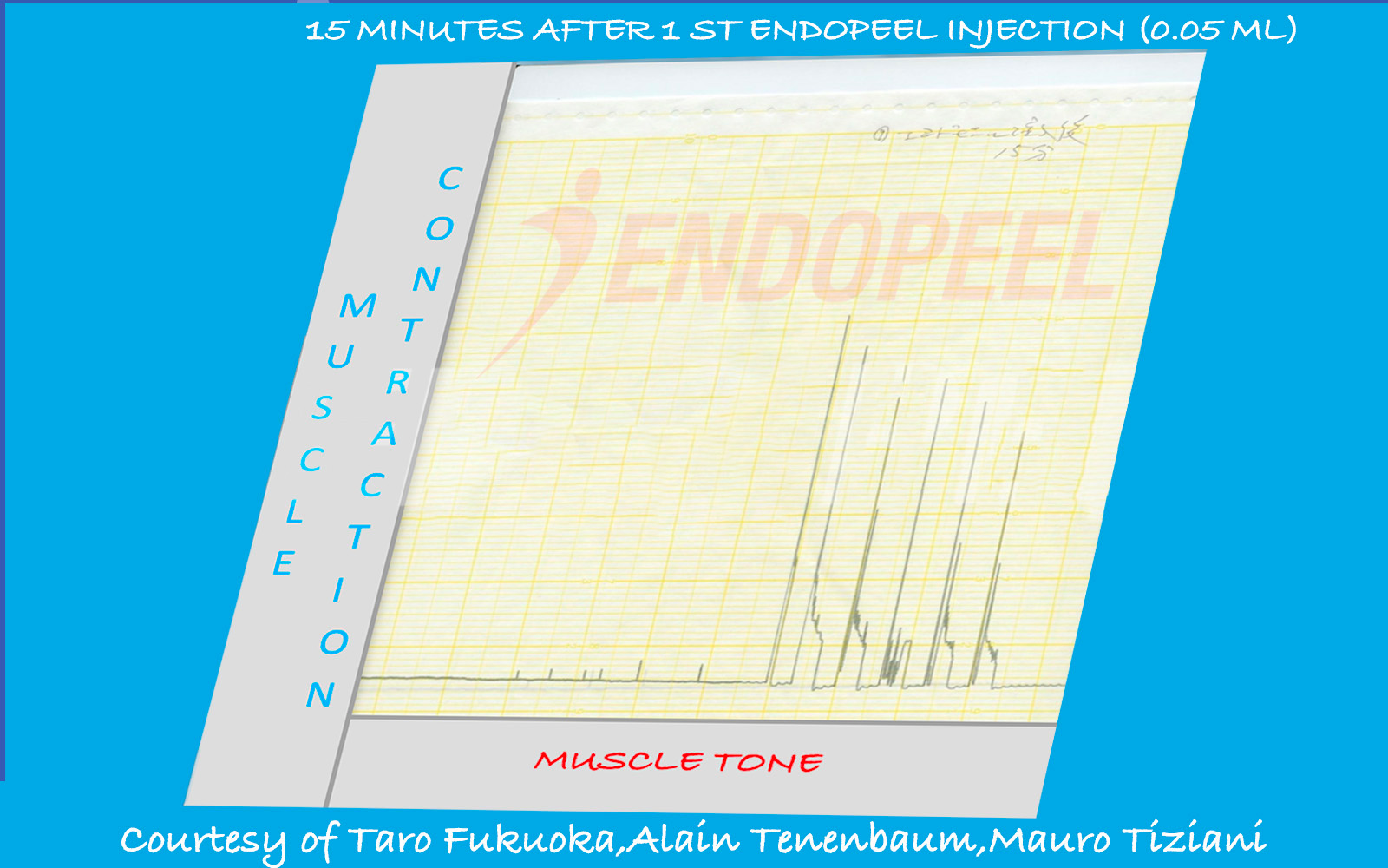
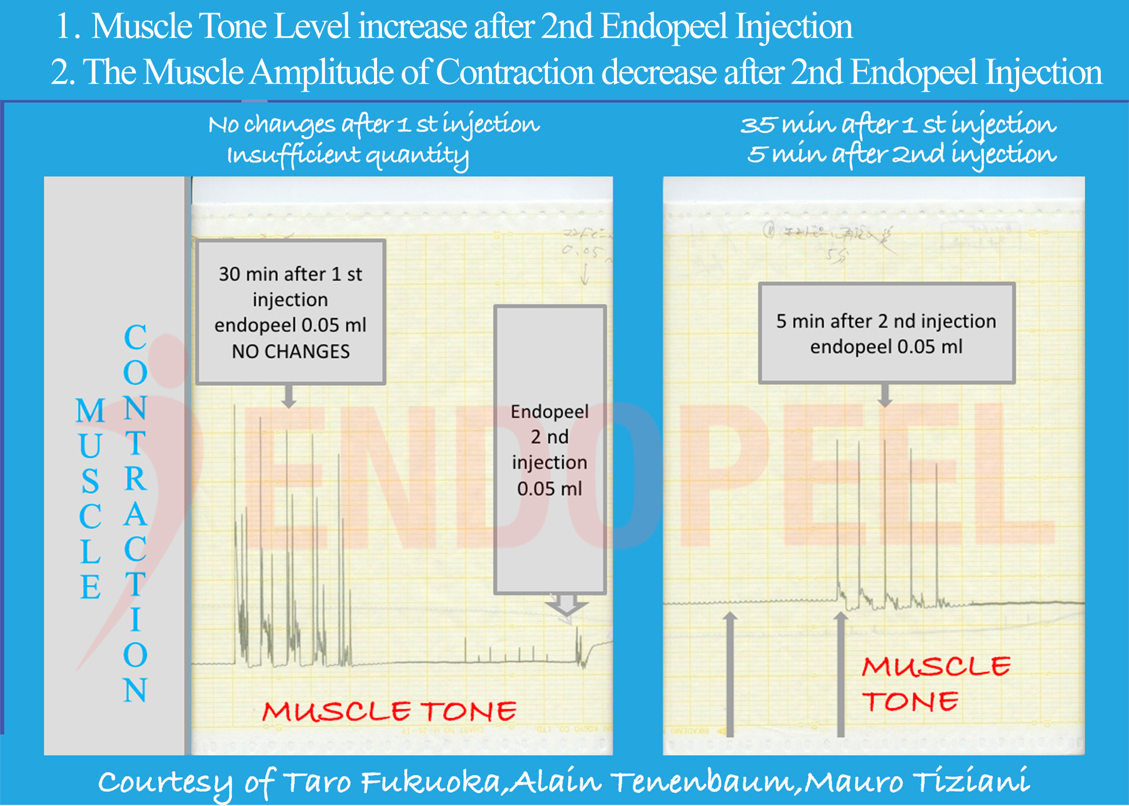
This page presents the physiologic foundation of Endopeel® based on experimental muscle contraction studies conducted at Akita University School of Medicine in collaboration with the Kyoto Pathological Research Institute (2006–2007).

Endopeel® physiology experimental muscle contraction analysis

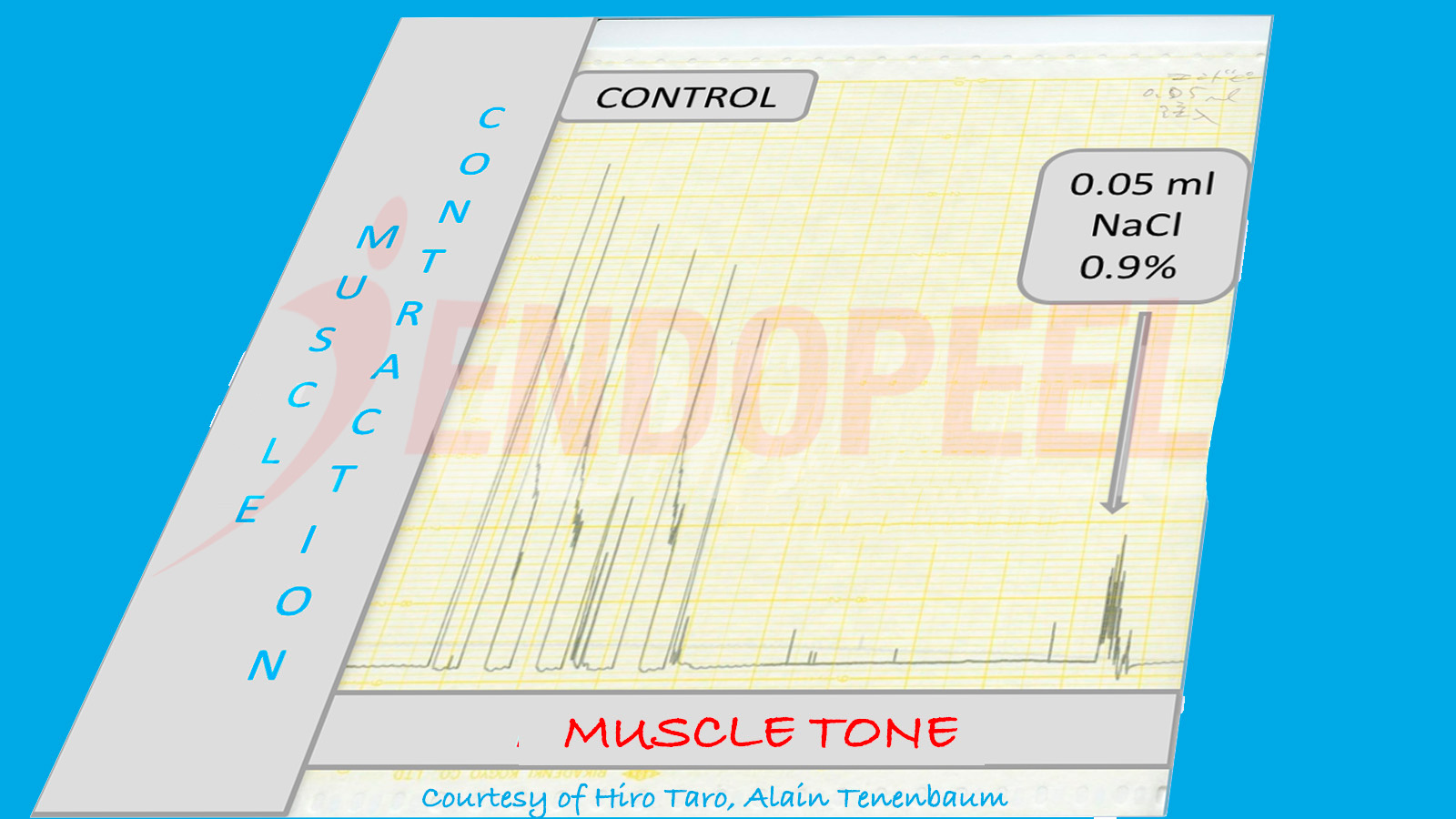
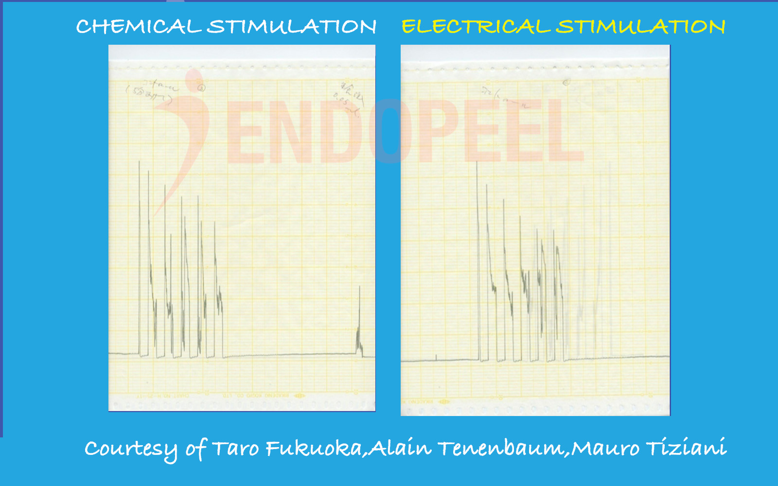
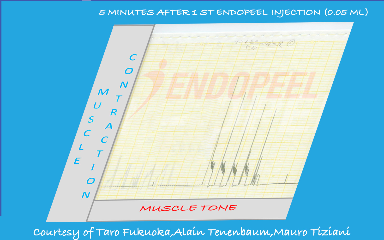
Data shown below are presented as physiologic tracings and comparative stimulation tests (electrical versus chemical), followed by the observed response after sequential Endopeel® injections.

Abstract

Physiologic Summary

This experimental physiologic study evaluated the effects of localized Endopeel® injection on muscle contraction amplitude and intrinsic muscle tone. Controlled measurements were performed using electrical and chemical stimulation protocols.

Results demonstrate reversible modulation of muscle contractility without neuromuscular blockade, denervation, or muscular atrophy. These findings support the classification of Endopeel® as a functional myomodulatory technique rather than a paralytic intervention.

Experimental model used to evaluate myotension (intrinsic muscle tone) and functional muscle behavior following Endopeel® injection
Figure 1. Experimental model used to record baseline tone and functional muscle response (myotension analysis).
Experimental Study

Akita University Physiologic Study

This experimental work (2006–2007) was conducted at Akita University School of Medicine in collaboration with the Kyoto Pathological Research Institute. The objective was to document changes in intrinsic muscle tone (myotension) and the resulting functional behavior of muscle tissue following localized Endopeel® administration.

Observed variations in apparent contraction amplitude are interpreted as a biomechanical consequence of increased tone and structural containment (myoplasty), rather than neuromuscular blockade. In clinical terms, this supports a functional triad: myotension (tone increase), myopexy (functional repositioning), and myoplasty (tissue tightening / confinement).

Authors
  • Prof. Dr. Hirotaro Fukuoka
  • Dr. Alain Tenenbaum, MD, PhD (Hon.)
  • Mr. Mauro Tiziani, Molecular Biologist; Red Cross Surgical Assistant (RCSA)
Baseline Recording

Baseline Muscle Tone and Functional Capacity

Prior to Endopeel® administration, baseline recordings were obtained to characterize the physiologic state of the muscle: resting tone (myotension) and overall functional responsiveness under standardized stimulation.

At baseline, the muscle demonstrated normal resting tonicity and unrestricted range of motion. This reference phase is essential, because subsequent reductions in apparent contraction amplitude can be interpreted correctly as a mechanical consequence of increased tone and structural confinement (myoplasty), rather than loss of neuromuscular function.

  • Baseline myotension: physiologic resting tone
  • Functional capacity: preserved responsiveness and movement
  • Interpretation framework: amplitude changes ≠ paralysis
Baseline gastrocnemius tracing illustrating physiologic resting tone (myotension) and normal functional response prior to Endopeel® injection
Figure 2. Baseline tracing: physiologic myotension and preserved functional responsiveness prior to injection.
Comparison of electrical stimulation versus chemical stimulation with NaCl 0.9% demonstrating preserved muscle responsiveness prior to Endopeel® injection
Figure 3. Electrical stimulation versus chemical stimulation (NaCl 0.9%): baseline muscle responsiveness prior to Endopeel®.
Pre-Injection Control Phase

Electrical vs Chemical Stimulation (NaCl 0.9%)

Before Endopeel® administration, the muscle was evaluated under two controlled activation methods: electrical stimulation and chemical stimulation with NaCl 0.9%.

The purpose of this comparison is to establish baseline functional integrity: the muscle remains responsive and viable, with physiologic resting tone (myotension) and reproducible activation patterns under both stimulation modalities.

  • Control stimulus: NaCl 0.9% (chemical)
  • Functional endpoint: preserved muscle responsiveness
  • Baseline framework: tone (myotension) and viability established prior to Endopeel®
Early Physiologic Response

5 Minutes After First Endopeel® Injection (0.05 mL – Single Point)

In this experimental phase, only 0.05 mL was injected at a single intramuscular point. This volume is deliberately minimal and primarily intended to observe early physiologic changes.

At this dose and single-point distribution, measurable increase in global muscle tone (myotension) may remain subtle and not yet produce a visible structural effect.

  • Injected volume lower than typical clinical application
  • Single injection point only
  • Dose-dependent effect expected

Clinically, structural myotension effects are achieved either through multiple distributed injections (0.05–0.1 mL per point) or, when targeting a single confined area such as a trigger zone, through higher localized volume (up to approximately 1 mL).

Muscle tracing five minutes after first 0.05 mL Endopeel® injection at a single point
Figure 4. Early physiologic response after minimal single-point injection (0.05 mL).
Muscle tracing fifteen minutes after Endopeel® injection demonstrating biomechanical confinement with preserved vitality
Figure 5. Functional adjustment within a progressively confined structural environment.
Structural Adaptation Phase

15 Minutes Post Injection: Myotension and Myoplasty

Fifteen minutes after injection, intrinsic muscle tone (myotension) remains elevated. The muscle progressively adapts to a more compact structural configuration.

Apparent reduction in contraction amplitude at this stage does not indicate neuromuscular inhibition. Instead, it reflects mechanical confinement within a tightened structural envelope — the beginning of the myoplasty effect.

Biomechanical analogy: A muscle operating freely within a large anatomical space can exhibit full excursion. When the same muscle is structurally confined within a smaller environment, its movement remains possible but its amplitude is naturally reduced — without paralysis and without loss of vitality.

  • Myotension remains increased
  • Functional movement preserved
  • Amplitude adjustment is biomechanical, not neurotoxic
Reinforcement Phase

Second Injection: Myotension Reinforcement and Myopexy

Following the second localized Endopeel® injection, intrinsic muscle tone demonstrates further reinforcement. The structural tightening effect becomes more pronounced.

This cumulative increase in myotension promotes functional repositioning of the muscle within its anatomical compartment — the basis of the myopexy effect.

As structural confinement intensifies (myoplasty), movement range adapts naturally. The muscle remains viable and contractile, yet operates within a more compact and stabilized framework.

  • Reinforced intrinsic tone (myotension)
  • Functional repositioning (myopexy)
  • Structural containment (myoplasty)
Muscle tracing following second Endopeel® injection demonstrating reinforced myotension and structural stabilization
Figure 6. Reinforced myotension and progressive structural stabilization after second injection.
Conclusion

Functional Myotension Without Paralysis

The experimental findings demonstrate that Endopeel® does not induce neuromuscular blockade. Instead, its primary mechanism is an increase in intrinsic muscle tone — myotension.

Progressive tone reinforcement produces structural tightening of the muscular compartment (myoplasty), leading to functional repositioning of the muscle within its anatomical environment (myopexy).

Apparent reductions in contraction amplitude observed in the experimental model reflect biomechanical confinement, not paralysis. Muscle vitality and functional capacity remain preserved.

These experimental observations describe physiologic behavior and should be interpreted within the context of the experimental model.

FAQ