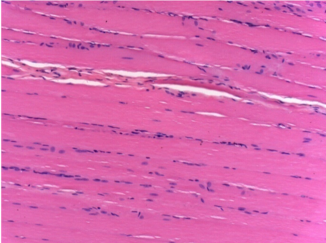
Histology

On Wistar rats

Courtesy of Lab of Pathology Department on Medicine School of Ribeirão Preto – USP
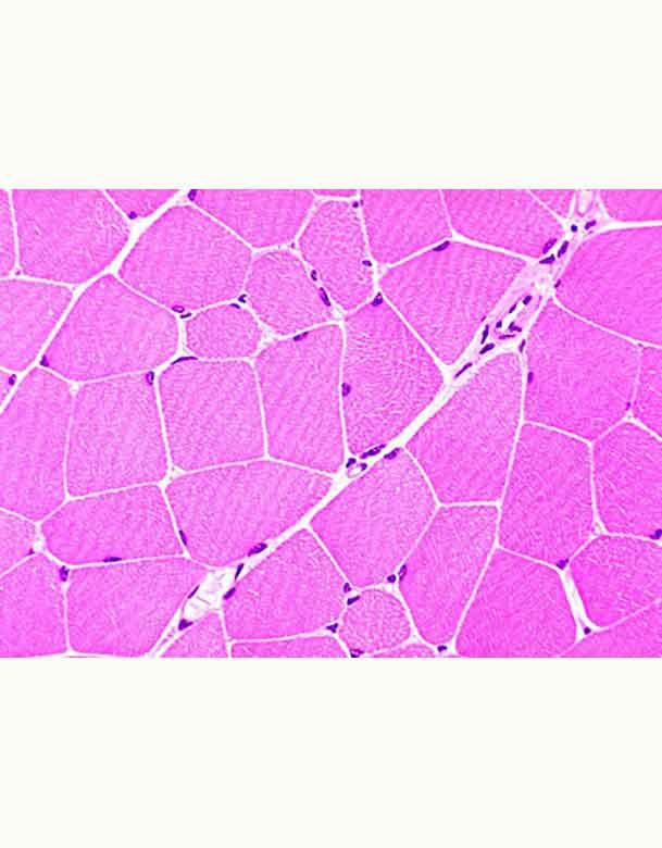
histology

01

Study
Group

Experimentators : R.Mené,M.Kim,A.Tenenbaum

N= 29 Wistar rats
Weight mean : 421g

Origin of animals : Biotério Central of Campus of Ribeirão Preto of São Paulo University
Maintenance : Experimental Pathology Lab of Pathology Department on Medicine School of Ribeirão Preto – USP
Animals keeping : polypropylen boxes
Animals food : basic diet of lab and water
Light conditions : natural light/dark cycles under controlled basic conditions
Experimentators : A.Tenenbaum,R.Mené,Kim

pathology department

02

Animals Sacrifice & Histologic Samples

Conventional Microscopy

The mice were euthanized in CO2 chambers.

  • Incision of the skin with a blade nº 23
  • Finding and isolation of the pretibial muscle using Metzenbaum scissors
  • Section of tendons with a Mayo scissors.
  • Fixation of Portions of the muscles on sections of cork oak
  • Embedding using 50% formaldehyde solution - 10% buffered and 50% of pure paraffin for conventional optical microscopic study.
  • Staining Sections as routine histological process.
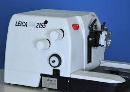
  • Microtome used : Leika RM 2155
  • Coloration of muscle slices with

-hematoxilin-eosin
-Masson´s trichrome
-and red picro-sirius.
  • Histologic examination was realized with a conventional microscope (Olympus BH-2).
  • Observation under polarized lampe
  • Photographic documentation was realized with a Leika DMR microscope joined to a digital camera Leika CD 300F and compatible PC.
microtome

03

Material

6 rats/box
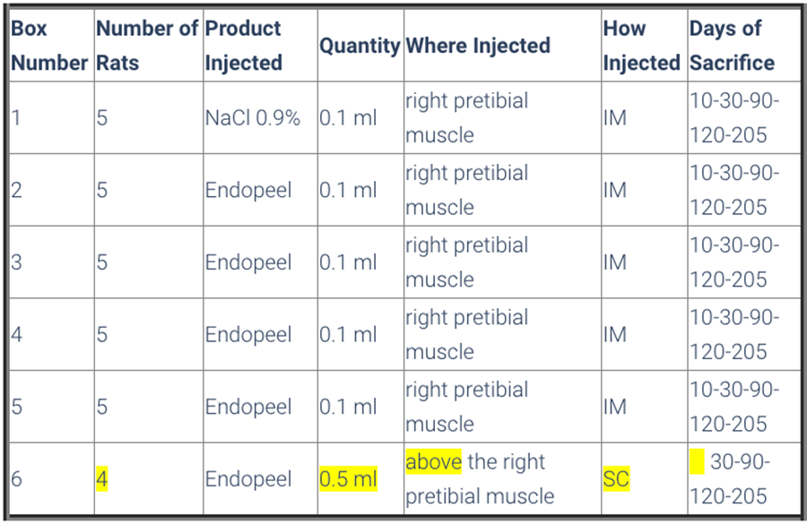
  • Animals were divided in 6 boxes, so that boxes from number 1 to 5 had 5 rats and box number 6 had 4 rats 
  • All animals were submitted to a walking evaluation by footprint impression on special paper , and previous immersion of back legs on a soap solution. 
  • Box 1 had the control group, that received the application of 0,1 ml saline solution ( NaCl 0,9%) on the right pretibial muscle. 
  • The mice of boxes 2 to 5 received 0,1 ml of Endopeels Original Main Product on the same muscle as control group.

  • Box 6 was constituted of 4 rats, and each one received 0,5 ml of Endopeels original main product injected on the subcutaneous layer. 

  • All the animals were submitted to walking evaluation before and after their respective injections.
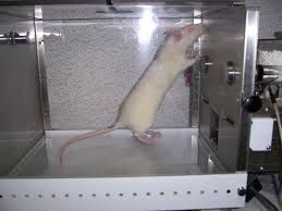
boxes for rats

04

Methods

Animals are sacrified after 10 days-1 month- 3 months-7 months
sacrifice table

05

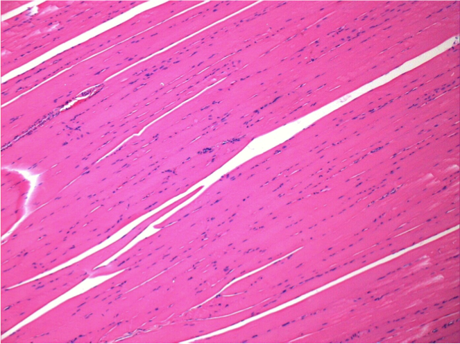
Control

After injection of NaCl 0.5%

Comment : Nothing to declare after saline solution injection
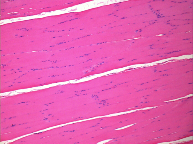
without treatment

L:Pretibial-No treatment

R:Pretibial-After 0.1 ml NaCl 0.9% IM

06

10 days after Endopeel Injection

Box 2

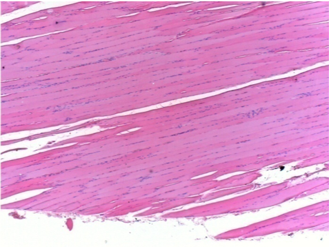
10 days after Endopeel Injection 0.1ml in the right pretibial muscle.

Here you may see the formation of the vacuoles which are surrounded by lymphocytes. Vacuoles are different from tissue necrosis . The presence of lymphocytes is related to the permeability of the cell membranes.


left control- 100xD10

L : Control-100xD10

right 100xD10

R:100xD10

right200xx

R :200xD10

right400xxD10

R :400xD10

07

1 month after Endopeel Injection

Box 3

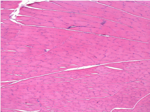
1 month after Endopeel Injection 0.1ml in the right pretibial muscle.
What is seen in black on the pictures is not a necrosis like could imagine some scientifics !
In fact, 4 conclusions have to be taken in consideration

  • an artefact of coloration
  • an absence of necrosis
  • an apoptosis
  • a bioregenerative process 
left control- 100xD30

L : Control-100xD30

right 100x

R:100xD30

right400xxD30

R :400xD30

08

3 months after Endopeel Injection

Box 4

3 months (D90)after Endopeel Injection 0.1ml in the right pretibial muscle.

left control- 100xD90

L : Control-100xD90

right 100x

R:100xD90

09

7 months after Endopeel Injection

Box 5

7 months (D210)after Endopeel IM Injection 0.1ml in the right pretibial muscle.

Complete Restitutio ad integrum after 7 months

left control- 100xD210

L : Control-100xD210

right 100x

R:100xD210

L Control 50xD210

L :Control 50xD210

R50X-D210

R50X-D210


10

Endopeel Injection in Subcutaneous Tissue

Box 6

0.5 ml ( 5x 0.1ml)  Endopeel SC Injection in the right subcutaneous pretibial area.
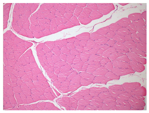
L:200x-Control-SC
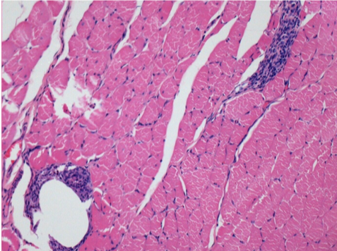
R-D10-SC-200X
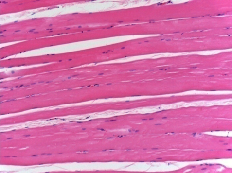
R-D10-SC-200X

R-D30-SC-200X

R-D30-SC-200X

R-D90-SC-200X
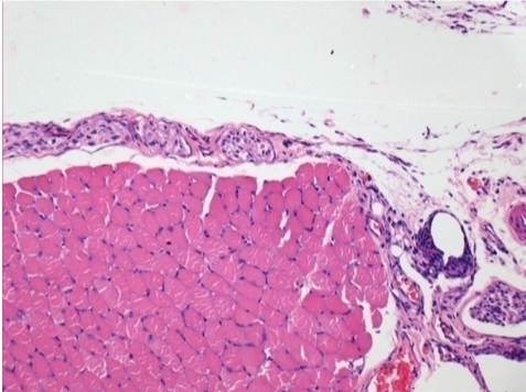
R-D90-SC-200X

R-D210-SC-200X
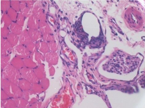
R-D210-SC-200X

R-D210-SC-400X

R-D210-SC-400X


conclusions
    • Endopeel induces a selective reversible myofibrolysis and  inflammatory reaction on a period of 1 month, approximately
    • Muscular changes are reversible in almost   full totality
    • The muscle is the better place to inject Endopeel because of more efficacity, control and duration of  its action
    • No necrosis nor abcess have been found all over the study.

Other Study applied to Gynecology

Other Study